Back Muscles Diagram : A P Back Muscles Diagram Quizlet. Another common cause of lower back and hip pain is disc injury. The human back extends from the buttocks to the posterior portion of the neck and shoulders. Superficial, intermediate, deep and deepest layers.these muscles lie on each side of the vertebral column, deep to the thoracolumbar fascia they span the entire length of the vertebral column, extending from the cranium to the pelvis Deep muscles of the lower back include: The deltoid, teres major, teres minor, infraspinatus, supraspinatus (not shown) and subscapularis muscles (not shown) all extend from the scapula to the humerus and act on the shoulder joint.
Deep back muscles superficial back muscles action movements of the shoulder. The part of the nerve that emerges out of the spine is called the nerve root. This muscle is a major generator of lower back and hip pain, as well as being responsible for complaints of a burning sensation along the posterior superior iliac spine (psis) and sacroiliac joint. The pelvis at the bottom of the back and the shoulders at the top of the back give the back. Lower back muscle diagram anatomy does degenerative disc disease affect the lower back muscle?
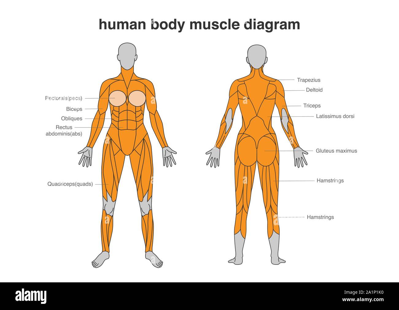
Muscles Of The Back Teachmeanatomy from teachmeanatomy.info Support and protect your spine; The part of the nerve that emerges out of the spine is called the nerve root. See back muscles and low back pain. The muscles of your back support your spine, attach your pelvis and shoulders to your trunk, and provide mobility and stability to your trunk and spine. The muscles of the lower back help stabilize, rotate, flex, and extend the spinal column, which is a bony tower of 24 vertebrae that gives the body structure and houses the spinal cord.the spinal. By sport fitness advisor staff. The muscles of the back that work together to support the spine, help keep the body upright and allow twist and bend in many directions. There are several different layers of muscles in your back that are often pulling in different and various directions.
Muscle strain is often the cause of back pain from heavy lifting or vigorous exercise.
Deep muscles of the lower back include: Lower back muscle diagram anatomy does degenerative disc disease affect the lower back muscle? Three types of back muscles that help the spine function are extensors, flexors and obliques. Some of the links in the post above are affiliate links.. On these diagrams of back muscle, you'll learn about back muscles, their locations and functional anatomy. Superficial, intermediate, deep and deepest layers.these muscles lie on each side of the vertebral column, deep to the thoracolumbar fascia they span the entire length of the vertebral column, extending from the cranium to the pelvis The back has a total of 40 muscles. For example, some muscles located in the chest also help move the shoulders. The muscles, bones, ligaments, and tendons in the back can all be injured and cause back pain. The muscles of the back can be arranged into 3 categories based on their location: These muscles include the large paired muscles in the lower back, called erector spinae, which help hold up the spine, and gluteal muscles. Likewise, there are muscles in other parts of the body that help support and move the spine. The deltoid, teres major, teres minor, infraspinatus, supraspinatus (not shown) and subscapularis muscles (not shown) all extend from the scapula to the humerus and act on the shoulder joint.
Most of the time, back muscle pain is diagnosed then treated with little more than a prescription of rest, painkillers and muscle relaxants. Superficial back muscles, intermediate back muscles and intrinsic back muscles.the intrinsic muscles are named as such because their embryological development begins in the back, oppose to the superficial and intermediate back muscles which develop elsewhere and are therefore classed as extrinsic muscles. Intermediate back muscles and c. The human back extends from the buttocks to the posterior portion of the neck and shoulders. The back has a total of 40 muscles.
Pin On Health from i.pinimg.com Related posts of back muscles chart muscle anatomy diagram. This is a diagram of the larger and more surface muscles of the low back. Superficial, intermediate, deep and deepest layers.these muscles lie on each side of the vertebral column, deep to the thoracolumbar fascia they span the entire length of the vertebral column, extending from the cranium to the pelvis Support and protect your spine; Anatomynote.com found anatomy of back muscles diagram from plenty of anatomical pictures on the internet. This muscular system diagram shows the major muscle groups from the back or posterior view. We think this is the most useful anatomy picture that you need. On these diagrams of back muscle, you'll learn about back muscles, their locations and functional anatomy.
On these diagrams of back muscle, you'll learn about back muscles, their locations and functional anatomy.
The muscles of your back support your spine, attach your pelvis and shoulders to your trunk, and provide mobility and stability to your trunk and spine. The back muscles can be three types. Back to tracking tools main page. The deep back muscles, also called intrinsic or true back muscles, consist of four layers of muscles: Some of the links in the post above are affiliate links.. This muscle is a major generator of lower back and hip pain, as well as being responsible for complaints of a burning sensation along the posterior superior iliac spine (psis) and sacroiliac joint. This is a diagram of the larger and more surface muscles of the low back. Creatine research more than a sports supplement read more…. Human muscle system, the muscles of the human body that work the skeletal system, that are under voluntary control, and that are concerned with movement, posture, and. On these diagrams of back muscle, you'll learn about back muscles, their locations and functional anatomy. Creatine is now proving to be one of the most potent muscle growth accelerators giving excellent muscle mass increase and phenomenal strength increases order yours today. We think this is the most useful anatomy picture that you need. Related posts of muscles of the lower back and buttocks diagram anatomy muscles view.
Superficial, intermediate, deep and deepest layers.these muscles lie on each side of the vertebral column, deep to the thoracolumbar fascia they span the entire length of the vertebral column, extending from the cranium to the pelvis How many muscles are in the back? The human back extends from the buttocks to the posterior portion of the neck and shoulders. Anatomynote.com found anatomy of back muscles diagram from plenty of anatomical pictures on the internet. The anatomy of your back muscles can be complex.
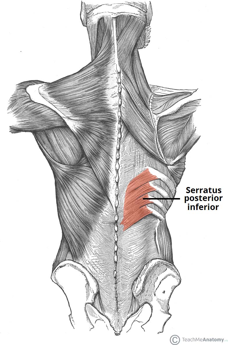
Woman Body Muscles Diagram In Full Length Front And Back Side Stock Vector Image Art Alamy from c8.alamy.com Superficial back muscles, intermediate back muscles and intrinsic back muscles.the intrinsic muscles are named as such because their embryological development begins in the back, oppose to the superficial and intermediate back muscles which develop elsewhere and are therefore classed as extrinsic muscles. These muscles include the large paired muscles in the lower back, called erector spinae, which help hold up the spine, and gluteal muscles. By sport fitness advisor staff. The anatomy of your back muscles can be complex. As you can see, there are also have a spine of scapula deltoid, triceps brachii, latissimus dorsi. How many muscles are in the back? Nerves in your lower back. Below you'll see diagrams along with the names of the back muscles that may be the cause of your pain.
Pain log more pain mapping tools
Below you'll see diagrams along with the names of the back muscles that may be the cause of your pain. Daniel nelson on january 1, 2019 2 comments 🔥! To learn more about the anatomy of the spine, watch this video. Some of the links in the post above are affiliate links.. People with back pain people who experience headaches printing for use during doctor visits to communicate information about your symptoms quickly tracking your progress over time related tools: Muscles of lower back diagram. We think this is the most useful anatomy picture that you need. The muscles of the lower back help stabilize, rotate, flex, and extend the spinal column, which is a bony tower of 24 vertebrae that gives the body structure and houses the spinal cord.the spinal. Pain log more pain mapping tools Support and protect your spine; The muscles, bones, ligaments, and tendons in the back can all be injured and cause back pain. For example, some muscles located in the chest also help move the shoulders. Both the deltoid and the trapezius are firmly attached to the spine of the scapula.
Berbagi :
Posting Komentar
untuk "Back Muscles Diagram : A P Back Muscles Diagram Quizlet"


Posting Komentar untuk "Back Muscles Diagram : A P Back Muscles Diagram Quizlet"